Fetal Wound Healing and Translational Applications for Scarless Skin Repair
DOI:
https://doi.org/10.22034/JATE.2026.224Keywords:
Wound healing, Scarless, Translational application, TGFBAbstract
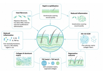
Fetal wound healing represents a unique model of scarless tissue repair and provides an important biological framework for developing regenerative therapies for adult skin wounds. Unlike adult wound healing, which often results in fibrosis and permanent scar formation, early-gestation fetal wounds can restore normal skin architecture with minimal inflammation, rapid re-epithelialization, organized extracellular matrix deposition, and reduced myofibroblast activation. This review summarizes the cellular, molecular, immunological, and biomechanical mechanisms underlying fetal scarless wound healing and discusses their translational relevance for tissue engineering and regenerative medicine. Key features of fetal repair include a hyaluronic acid-rich extracellular matrix, a higher collagen III/I ratio, increased transforming growth factor-β3 activity relative to TGF-β1 and TGF-β2, attenuated inflammatory cytokine signaling, fetal-specific fibroblast phenotypes, and a mechanically compliant wound environment. Particular attention is given to mechanotransduction pathways, including integrin-mediated signaling, focal adhesion kinase, Rho/ROCK, and YAP/TAZ, which link matrix stiffness and tissue tension to fibroblast activation and scar formation. The review further examines fetal-inspired therapeutic strategies, including hyaluronic acid-based hydrogels, immunomodulatory biomaterials, stem cell-derived secretomes, controlled growth factor delivery, anti-fibrotic agents, RNA-based therapies, and bioengineered skin substitutes. Although these approaches have shown promising preclinical and early clinical potential, true scarless regeneration in adult human skin remains challenging due to the complexity of the fetal wound microenvironment, differences in immune maturity, delivery timing, safety concerns, and regulatory barriers. Understanding fetal wound healing as an integrated and dynamically regulated process may guide the design of multi-cue regenerative therapies aimed not only at reducing scar formation but also at restoring functional skin architecture.
References
Weng T, Zhang L, Liu Y, Zhao X, Yue X. Advances in Tissue Engineering and Biomaterials for Minimizing Wound Scarring: Current Status and Future Challenges. Journal of Functional Biomaterials. 2025;17(1):3.
Prakoeswa FRS, Dzikri DNa. Scarless Wound Healing: Recent Advances and Innovations. Cermin Dunia Kedokteran. 2025;52(8):546–52.
Mascharak S, desJardins-Park HE, Davitt MF, Guardino NJ, Gurtner GC, Wan DC, et al. Modulating cellular responses to mechanical forces to promote wound regeneration. Advances in Wound Care. 2022;11(9):479–95.
Niti A, Kouzi-Koliakou K, Michopoulou A. Fibroblast Lineage Switching as the Developmental Origin of Scarring and Target for Regenerative Healing. Biology. 2026;15(5):409.
Namazi MR, Fallahzadeh MK, Schwartz RA. Strategies for prevention of scars: what can we learn from fetal skin? International journal of dermatology. 2011;50(1):85–93.
Buhimschi CS, Dulay AT, Abdel‐Razeq S, Zhao G, Lee S, Hodgson EJ, et al. Fetal inflammatory response in women with proteomic biomarkers characteristic of intra‐amniotic inflammation and preterm birth. BJOG: An International Journal of Obstetrics & Gynaecology. 2009;116(2):257–67.
Mohsin S, Prinindya KNN, Nawshad M, Hasan A, Augustine R, Mahdi E. 3D Bioprinting Strategies for Diabetic Wound Healing: Functional Bioinks, Vascularization, and Immune Modulation. Bioprinting. 2026:e00486.
Chakrabarti S, Ahmed RK, Nath S, Nandi A, Pathak MP. Scar-Free Wound Healing: Advances in Biomaterial-Based Formulations, Smart Dressings and Bioactive Therapeutics–A Comprehensive Review. Biochemical and Biophysical Research Communications. 2026:153839.
Satish L, Kathju S. Cellular and molecular characteristics of scarless versus fibrotic wound healing. Dermatology research and practice. 2010;2010(1):790234.
Parekh A, Hebda PA. The contractile phenotype of dermal fetal fibroblasts in scarless wound healing. Current pathobiology reports. 2017;5(3):271–7.
Beanes SR, Hu F-Y, Soo C, Dang CM, Urata M, Ting K, et al. Confocal microscopic analysis of scarless repair in the fetal rat: defining the transition. Plastic and reconstructive surgery. 2002;109(1):160–70.
Buchanan EP, Longaker MT, Lorenz HP. Fetal skin wound healing. Advances in clinical chemistry. 2009;48:137–61.
Rolfe K, Grobbelaar A. A review of fetal scarless healing. International Scholarly Research Notices. 2012;2012(1):698034.
Singampalli KL, Balaji S, Wang X, Parikh UM, Kaul A, Gilley J, et al. The role of an IL-10/hyaluronan axis in dermal wound healing. Frontiers in cell and developmental biology. 2020;8:636.
Zgheib C, Xu J, Liechty KW. Targeting inflammatory cytokines and extracellular matrix composition to promote wound regeneration. Advances in wound care. 2014;3(4):344–55.
Zhao F, Lang H, Wang Z, Zhang T, Zhang D, Wang R, et al. Human novel microRNA Seq-915_x4024 in keratinocytes contributes to skin regeneration by suppressing scar formation. Molecular Therapy Nucleic Acids. 2019;14:410–23.
Zheng Z, Zhang X, Dang C, Beanes S, Chang GX, Chen Y, et al. Fibromodulin is essential for fetal-type scarless cutaneous wound healing. The American Journal of Pathology. 2016;186(11):2824–32.
Xia Z-Y, Wang Y, Shi N, Lu M-Q, Deng Y-X, Qi Y-J, et al. Fetal mice dermal mesenchymal stem cells promote wound healing by inducing M2 type macrophage polarization. World Journal of Stem Cells. 2025;17(2):101030.
Balaji S, King A, Marsh E, LeSaint M, Bhattacharya SS, Han N, et al. The role of interleukin-10 and hyaluronan in murine fetal fibroblast function in vitro: implications for recapitulating fetal regenerative wound healing. PLoS One. 2015;10(5):e0124302.
Su C-Y, Liu T-Y, Wang H-V, Yang W-C. Histopathological Study on Collagen in Full-Thickness Wound Healing in Fraser’s Dolphins (Lagenodelphis hosei). Animals. 2023;13(10).
Dang CM, Beanes SR, Lee H, Zhang X, Soo C, Ting K. Scarless fetal wounds are associated with an increased matrix metalloproteinase–to–tissue-derived inhibitor of metalloproteinase ratio. Plastic and reconstructive surgery. 2003;111(7):2273–85.
Jerrell RJ, Leih MJ, Parekh A. The altered mechanical phenotype of fetal fibroblasts hinders myofibroblast differentiation. Wound Repair and Regeneration. 2019;27(1):29–38.
Hsu M, Peled ZM, Chin GS, Liu W, Longaker MT. Ontogeny of expression of transforming growth factor-β1 (TGF-β1), TGF-β3, and TGF-β receptors I and II in fetal rat fibroblasts and skin. Plastic and reconstructive surgery. 2001;107(7):1787–94.
Zheng Z, James AW, Li C, Jiang W, Wang JZ, Chang GX, et al. Fibromodulin reduces scar formation in adult cutaneous wounds by eliciting a fetal-like phenotype. Signal transduction and targeted therapy. 2017;2(1):17050.
Li H, Luo S, Li H, Pan H, Jiang L, Chen Y, et al. From fetal tendon regeneration to adult therapeutic modalities: TGF-β3 in scarless healing. Regenerative Medicine. 2023;18(10):809–22.
Geng Y, Zhang W, Dun X, Wang Y, Shao Y. Rapid cutaneous wound healing in nude mice by fetal skin-derived stem cells involves enhanced collagen secretion and angiogenesis. Frontiers in Medicine. 2025;12:1557973.
Brant JO, Lopez M-C, Baker HV, Barbazuk WB, Maden M. A comparative analysis of gene expression profiles during skin regeneration in Mus and Acomys. PLoS one. 2015;10(11):e0142931.
Takaya K, Sunohara A, Aramaki-Hattori N, Sakai S, Okabe K, Kanazawa H, et al. Role of Wnt signaling in mouse fetal skin wound healing. Biomedicines. 2022;10(7):1536.
Wang X, Jiao Y, Pan Y, Zhang L, Gong H, Qi Y, et al. Fetal dermal mesenchymal stem cell‐derived exosomes accelerate cutaneous wound healing by activating notch signaling. Stem cells international. 2019;2019(1):2402916.
Takaya K, Aramaki-Hattori N, Sakai S, Okabe K, Kishi K. Effect of sonic hedgehog on the regeneration of epidermal texture patterns. Biomedicines. 2022;10(12):3099.
Podolak-Popinigis J, Ronowicz A, Dmochowska M, Jakubiak A, Sachadyn P. The methylome and transcriptome of fetal skin: implications for scarless healing. Epigenomics. 2016;8(10):1331–45.
Cheng J, Yu H, Deng S, Shen G. MicroRNA profiling in mid-and late-gestational fetal skin: implication for scarless wound healing. The Tohoku journal of experimental medicine. 2010;221(3):203–9.
Lang H, Zhao F, Zhang T, Liu X, Wang Z, Wang R, et al. MicroRNA-149 contributes to scarless wound healing by attenuating inflammatory response. Molecular Medicine Reports. 2017;16(2):2156–62.
Patten J, Albeltagy N, Bonadio JD, Ortez A, Wang K. FAK Differentially Mechanoregulates Cell Migration during Wound Closure. ACS biomaterials science & engineering. 2025;11(8):4968–77.
Zhu C, Li L, Zhao B. The regulation and function of YAP transcription co-activator. Acta Biochim Biophys Sin. 2015;47(1):16–28.
Veerasubramanian PK. Microenvironmental cues epigenetically modulate macrophage inflammation: University of California, Irvine; 2022.
Short WD, Parikh UM, Templeman N, Olutoye OO, Blum A, Colchado D, et al. Phenotypic Differences in Adult and Fetal Dermal Fibroblast Responses to Mechanical Tension. bioRxiv. 2020:2020.09. 30.319749.
Lin H, Wang X, Chung M, Cai S, Pan Y. Direct fibroblast reprogramming: an emerging strategy for treating organic fibrosis. Journal of Translational Medicine. 2025;23(1):240.
Walraven M, Talhout W, Beelen RH, van Egmond M, Ulrich MM. Healthy human second‐trimester fetal skin is deficient in leukocytes and associated homing chemokines. Wound Repair and Regeneration. 2016;24(3):533–41.
Liechty KW, Kim HB, Adzick NS, Crombleholme TM. Fetal wound repair results in scar formation in interleukin-10–deficient mice in a syngeneic murine model of scarless fetal wound repair. Journal of pediatric surgery. 2000;35(6):866–73.
Roy A, Mantay M, Brannan C, Griffiths S. Placental tissues as biomaterials in regenerative medicine. BioMed research international. 2022;2022(1):6751456.
Gansevoort M, Oostendorp C, Bouwman LF, Tiemessen DM, Geutjes PJ, Feitz WF, et al. Collagen-heparin-FGF2-VEGF scaffolds induce a regenerative gene expression profile in a fetal sheep wound model. Tissue Engineering and Regenerative Medicine. 2024;21(8):1173–87.
Avila-Martinez N, Pfirrmann M, Gomes ML, Krymchenko R, Versteeg EM, Vlig M, et al. Effect of Hyaluronan in Collagen Biomaterials on Human Macrophages and Fibroblasts In Vitro. Journal of Functional Biomaterials. 2025;16(5):167.
Rong H, Dong Y, Zhao J, Zhang X, Li S, Sun Y, et al. Fetal milieu-simulating hyaluronic acid-dopamine-chondroitin sulfate hydrogel promoting angiogenesis and hair regeneration for wound healing. International Journal of Biological Macromolecules. 2023;248:125739.
Esfahlani MA, Ebrahimi-Barough S, Imani R, Lotfibakhshaiesh N, Bahrami N, Namini MS, et al. Curcumin nanoparticles and mesenchymal stem cell exosomes embedded in gelatin hydrogel enhance healing of severe burns. Burns. 2026:107990.
Huang L, Lin Z, Liu H, Lin X, Liao N, Wu X. Mesenchymal stem cell-conditioned medium accelerates type 2 diabetic wound healing by targeting TNF and chemokine signaling. Frontiers in Cell and Developmental Biology. 2025;13:1659444.
Jiang D, Correa-Gallegos D, Christ S, Stefanska A, Liu J, Ramesh P, et al. Two succeeding fibroblastic lineages drive dermal development and the transition from regeneration to scarring. Nature cell biology. 2018;20(4):422–31.
Lichtman MK, Otero‐Vinas M, Falanga V. Transforming growth factor beta (TGF‐β) isoforms in wound healing and fibrosis. Wound repair and regeneration. 2016;24(2):215–22.
Soo C, Beanes SR, Hu F-Y, Zhang X, Dang C, Chang G, et al. Ontogenetic transition in fetal wound transforming growth factor-β regulation correlates with collagen organization. The American journal of pathology. 2003;163(6):2459–76.
Colwell AS, Krummel TM, Longaker MT, Lorenz HP. Fetal and adult fibroblasts have similar TGF-β–Mediated, smad-dependent signaling pathways. Plastic and Reconstructive Surgery. 2006;117(7):2277–83.
Marchioni A, Tonelli R, Andreani A, Cappiello GF, Fermi M, Trentacosti F, et al. Molecular mechanisms and physiological changes behind benign tracheal and subglottic stenosis in adults. International journal of molecular sciences. 2022;23(5):2421.
Tacheau C, Michel L, Farge D, Mauviel A, Verrecchia F. Involvement of ERK signaling in halofuginone-driven inhibition of fibroblast ability to contract collagen lattices. European journal of pharmacology. 2007;573(1-3):65–9.
Leong EK, Al Bitar H, Marshall J, Bezuhly M. 28. Ketotifen Inhibits Dermal Fibroblast Responses To The Pro-fibrotic Effects Of TGF-beta. Plastic and Reconstructive Surgery–Global Open. 2024;12(S4):20.
Murali R, Balasubramanian RV, Kasoju N, NS R, Kartha RS, Kumar A, et al. Unravelling the wound healing efficiency of 3D bioprinted alginate-gelatin-diethylaminoethyl cellulose-fibrinogen based skin construct in a rat's full thickness wound model. International Journal of Biological Macromolecules. 2025;305:140816.
Song S, Guo J, Kong B, Liu R, Wang J, Lu L, et al. Black phosphorus-loaded anisotropic nanofiber sponges for random skin flaps regeneration. Chemical Engineering Journal. 2024;502:158133.
Meseci E, Kayatas S, Api M, Boza A, Cikman MS. Comparison of the effectiveness of topical silicone gel and corticosteroid cream on the pfannenstiel scar prevention—a randomized controlled trial. Ginekologia polska. 2017;88(11):591–8.
Mohamed SH, Omar GA-E, Hamdy AE-S. Microneedling with topical insulin versus microneedling with non-cross-linked hyaluronic acid for atrophic post-acne scars: a split-face study. Dermatology Practical & Conceptual. 2024;14(3):e2024183.
Ono I, Akasaka Y, Kikuchi R, Sakemoto A, Kamiya T, Yamashita T, et al. Basic fibroblast growth factor reduces scar formation in acute incisional wounds. Wound Repair and Regeneration. 2007;15(5):617–23.
Yang M, Yang Y, Shi G, Guo Y, Xuan T, Sun D, et al. Targeting Inflammatory and Fibrotic Responses: Butorphanol Orchestrates Wound Healing by Suppressing the IL-6/p38/JNK Pathway. DISCOVERY MEDICINE. 2025;37(201):2428–38.
Shi J, Zhang S, Zhang Z, Xu J, Chen Y, Sun S. Efficacy of triamcinolone acetonide combined with botulinum toxin A in the treatment of hypertrophic scars and keloids: A meta-analysis. Burns. 2024;50(9):107250.
Meuli M, Schiestl C, Hartmann-Fritsch F, Middelkoop E, Kim B-S, Neuhaus K, et al. Safety and efficacy of bio-engineered, autologous dermo-epidermal skin grafts in adolescent and adult burn patients: 1-year results of a prospective, randomized, controlled, multicenter phase IIB clinical trial. EClinicalMedicine. 2025;90.
Holmes IV JH, Gibson AL, Short T, Joe VC, Litt J, Carson J, et al. A phase 3b, open-label, single-arm, multicenter, expanded-access study of the safety and clinical outcomes of StrataGraft® treatment in adults with deep partial-thickness thermal burns. Burns. 2024;50(8):2013–22.
Allanki S, Strilic B, Scheinberger L, Onderwater YL, Marks A, Günther S, et al. Interleukin-11 signaling promotes cellular reprogramming and limits fibrotic scarring during tissue regeneration. Science advances. 2021;7(37):eabg6497.
Park J, Lee S, Chung J-Y, Yun S, Park HO, Park JY, et al. Safety and Pharmacokinetics of SRN001, a Novel siRNA Drug Targeting Amphiregulin via the SAMiRNA Platform. Drug Design, Development and Therapy. 2026:589997.
Downloads
Published
How to Cite
Issue
Section
License
Copyright (c) 2026 The Journal of Applied Tissue Engineering

This work is licensed under a Creative Commons Attribution-NonCommercial-NoDerivatives 4.0 International License.
